>>
Industry>>
Healthcare>>
How Spinal Imaging Technology ...How Spinal Imaging Technology Evolved Over the Last 130 Years
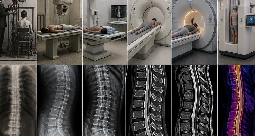
Before 1895, a physician who suspected a spine problem worked with two things: hands and judgment. Someone arrived with back pain that had kept them from working. The doctor pressed along the vertebrae, watched how the patient moved, asked where it hurt worst, then made a call based on experience alone. Whatever sat under the skin stayed there. Bone was invisible in a living body, and no technique existed to change that. Then Wilhelm Röntgen noticed a coated screen glowing on a bench several feet from the tube he was working with, and the practice of medicine changed permanently.
What followed reads less like steady progress than a string of lucky breaks. Nobel Prizes came into it. So did Cold War physics, and a quantity of mathematics borrowed from fields that had nothing to do with patients. The span runs 130 years and covers four separate technologies, moving from faint shadow images on glass plates to software that reviews thousands of lumbar studies between one shift and the next. Anyone who has had a scan of their back has watched the end of a very long chain of events.
The Accident That Changed Everything
Röntgen was working late in his Würzburg laboratory on November 8, 1895, running current through a cathode-ray tube he had wrapped in black cardboard. A screen coated with barium platinocyanide sat well across the room, far outside the reach of any cathode ray, and it was glowing. He stayed with the problem for weeks and barely left the building. Something coming off that tube passed through skin and muscle without slowing, then stopped cold at bone.
His wife Bertha held her hand in the beam, and the plate came back showing finger bones and her wedding ring. Hospitals across Europe were trying the method on patients within weeks of his first paper. By the close of 1896, more than 1,000 books and articles on X-rays had reached print, an uptake no medical technology has matched since. The 1901 Nobel Prize in Physics, the first ever awarded, went to Röntgen for the discovery. He handed the money to his university and took out no patents on any of it.
The spine gained and lost in the same instant. Cracked vertebrae, dislocated joints, and worn bone edges could finally be seen and measured. The discs sitting between them could not. Soft tissue gave the beam nothing to stop against, so the structures behind most back pain stayed blank on the film. That gap held for another 75 years.
CT Scanning: The Spine Gets Sliced
Computed tomography reached patients in the early 1970s, and for bone it marked the largest shift since Röntgen. Godfrey Hounsfield built the first clinical scanner at EMI, the company then earning most of its money from records. The machine rotates an X-ray source around the body and collects hundreds of readings from different angles. Software assembles those readings into slices, using reconstruction mathematics that Allan Cormack had worked out separately during the 1960s. The two shared a Nobel Prize in 1979.
Spine work changed with it. Canal width could be measured directly. Bony spurs pressing against a nerve root showed up clearly enough to plan an operation around. Vertebrae became readable in a way flat film never allowed. Discs did not follow. A herniated disc sits between two vertebrae and consists mostly of water and fibrocartilage, so it registers faintly at best on CT. What a scan showed was the bone's response to the problem rather than the problem itself. Reading a spine that way meant studying the shelf and guessing at the book.
MRI: Soft Tissue Finally Has a Voice
Magnetic resonance imaging reached what neither X-ray nor CT could touch. A strong magnetic field aligns hydrogen nuclei throughout the body's water, a radiofrequency pulse knocks them out of alignment, and the signal they release as they settle back gets converted into an image. Tissue with more water returns a different signal than tissue with less, which is precisely why soft structures appear. Felix Bloch and Edward Purcell had described the underlying resonance separately in 1946 and shared a Nobel Prize in Physics for it in 1952. Turning that effect into a picture of a living body took Paul Lauterbur and Peter Mansfield, whose imaging methods earned them a Nobel Prize in 2003.
FONAR shipped the first commercial scanner in 1980. Spinal MRI became the standard for soft-tissue diagnosis within roughly ten years. Disc height, disc hydration, the direction of a bulge, the extent of a rupture: all of it became measurable. L3,L4, L5 disc herniation shows up as a soft-tissue protrusion that both earlier methods missed entirely, and MRI put it on record before anyone considered an incision.
Surgery changed as a result. Operations once opened on symptom patterns alone now began with the location already known. MRI also turned up disc bulges and degeneration in people reporting no pain at all, which made imaging findings something to weigh against the patient rather than treat on sight.
What the Imaging Boom Looks Like in Numbers
The commercial history that followed those scientific breakthroughs is staggering. The global medical imaging market size was valued at USD 43.5 billion in 2025 and is projected to grow from USD 45.5 billion in 2026 to USD 64.7 billion by 2033, at a compound annual growth rate of 5.1%, according to a 2026 report by Grand View Research. MRI represents the single largest segment of that market by revenue. The technology that began as a physics curiosity now underpins a multi-billion-dollar global industry.
That growth reflects something real: the demand for accurate, non-invasive diagnosis has never been higher, and imaging is the only tool that reliably delivers it. Every aging population cohort worldwide drives a corresponding rise in spine-related imaging, because disc degeneration is almost universal after a certain age.
AI Reads the Scans Now
The most recent chapter in spine imaging history is being written right now, and it involves machine learning. Radiologists have long known that two readers of the same lumbar MRI often reach different conclusions. AI is beginning to change that.
A study from Harvard Medical School highlighted a deep learning model that reviewed nine degenerative spinal conditions across a dataset of 55,000 lumbar spine MRI studies affiliated with Mass General Brigham, with the model achieving accuracy rates of 77% to 97% across all pathologies of interest, as presented at the 2024 Radiological Society of North America conference and reported by Sg2 Intelligence. That range of accuracy is comparable to experienced subspecialists, which is a genuine shift in what automated tools can do.
AI in spine imaging is not replacing radiologists yet. It is doing something more immediately useful: flagging urgent findings faster, reducing the backlog on routine reads, and building a layer of consistency that human interpretation, however skilled, cannot fully achieve at volume.
"AI applied to spine imaging shows potential to improve diagnostic accuracy and efficiency, bridge capacity gaps, and elevate patient care."- Sg2 Intelligence Clinical Strategy Practice, 2025
The SCAN Framework: Four Eras, Four Capabilities
One way to organize 130 years of imaging progress is what I'd call the SCAN framework. Each letter names the core capability that defined its era:
|
Era |
SCAN Capability |
Primary Technology |
What It Showed the Spine
|
|
1895 to 1960s |
Structure |
Plain X-ray |
Bone alignment, fractures, gross deformity |
|
1970s to 1980s |
Cross-section |
CT scanner |
Canal width, bony spurs, vertebral detail |
|
1980s to 2010s |
Anatomy (soft tissue) |
MRI |
Discs, nerves, ligaments, cord compression |
|
2015 to present |
Normalization |
AI-assisted MRI/CT |
Consistent, standardized pathology classification at scale |
The SCAN framework is not a clinical classification. It's a reader's guide to understanding why imaging reports from different decades describe the same spine in completely different terms. The technology changed what could be seen, which changed what got written down, which changed what got treated.
A Practical Takeaway for Anyone Reading an Imaging Report Today
If you're looking at a spinal imaging report and trying to make sense of it, a few orienting points help:
- X-ray findings describe bone. They say almost nothing about discs or nerves.
- CT findings add bony detail and can hint at disc pathology through secondary changes, but soft tissue stays limited.
- MRI is the study of record for any disc, nerve, or ligament question. If your symptoms involve radiating pain, numbness, or weakness, and no MRI has been done, the picture is incomplete.
- AI-assisted reads are increasingly common but should always be reviewed by a qualified clinician. The algorithm flags. The doctor interprets.
- Older reports may use terminology that has since been standardized. An "annular tear" in a 2005 report and a "high-intensity zone" in a 2025 report may describe the same finding.
The next time you slide into an MRI bore and hear that rhythmic clanging, you're sitting inside 130 years of accumulated physics, engineering, and clinical courage. Röntgen's fluorescent screen is in there somewhere. So is Felix Bloch's Nobel lecture, and a Harvard deep learning model trained on 55,000 scans. That's nothing. That's one of the most productive accidents in scientific history, still paying forward. Which part of that journey do you think matters most to the average patient walking into a spine clinic today?













Comments